Characterization of titanium surfaces for dental implants with inorganic contaminant
Link: https://www.scielo.br/scielo.php?script=sci_arttext&pid=S1806-83242005000200006
Marilia Garcia DinizI; Marco Antonio Santos PinheiroI; Antonio Carlos Canabarro Andrade JuniorII; Ricardo Guimarães FischerIII
IAdjunct Professors, Department of Mechanical Engineering
IIAssistant Professor, School of Dentistry
IIIFull Professor, School of Dentistry – State University of Rio de Janeiro
Brazilian Oral Research
Print version ISSN 1806-8324On-line version ISSN 1807-3107
Braz. oral res. vol.19 no.2 São Paulo Apr./June 2005
https://doi.org/10.1590/S1806-83242005000200006
ABSTRACT
The aim of this research was to characterize titanium surfaces blasted with aluminum oxide (Al2O3) particles using the KS 400 digital image processing program. Samples of grade II titanium plates were submitted to blasting processes using particles of Al2O3, and treated with a hydrofluoric acid-based solution. Three digital images from each sample surface were obtained using Scanning Electron Microscopy, and half-quantitative chemical analyses were subsequently performed using Electron Dispersive Spectroscopy (EDS). In addition, parameters related to the alumina phase, such as the concentration level, the area and perimeter of the particles and their circular form factor were measured using KS 400. The mechanical/chemical treatment caused depressions up to 10 µm on homogeneous surfaces. Although the chemical attack significantly removed the alumina phase, residual particles could still be identified by the EDS. The average area occupied by the alumina phase on the samples surfaces was 469.32 (± 284.98) µm2, the particle average perimeter was 81.61 (± 27.68) µm, and the mean circular form factor was 0.60 (± 0.05). Characterizing the titanium surface is essential in the evaluation of the material manufacturing process because the presence of residual aluminum particles may have deleterious effects on the formation of the osseous/implant tissue.
Descriptors: Titanium; Dental implants; Aluminum oxide; Image processing, computer-assisted.
RESUMO
O objetivo deste trabalho foi caracterizar parâmetros relativos a superfícies de titânio submetidas a jateamento com partículas de óxido de alumínio (alumina - Al2O3) por meio de análise e processamento digital de imagem no programa KS 400 (Carl Zeiss, Oberkochen, Alemanha). Amostras de titânio grau 2 foram jateadas com Al2O3 e submetidas a tratamento químico com soluções à base de ácido fluorídrico. Três imagens digitais das superfícies de amostras distintas foram obtidas em microscópio eletrônico de varredura. Análises químicas foram realizadas por espectroscopia por dispersão de energia (EDS). As imagens foram analisadas e processadas no KS 400 para a obtenção de parâmetros digitais e quantificação da fase residual da alumina, tais como área, perímetro das partículas e fator de forma circular. O tratamento mecânico/químico promoveu crateras de até 10 µm em uma superfície homogênea em relação ao controle (tratamento somente mecânico). O ataque químico removeu de maneira significativa a alumina, porém partículas residuais foram identificadas pela EDS. A área total ocupada pela alumina nas amostras experimentais foi de 469,32 (± 284,98) µm2. O perímetro médio das partículas foi de 81,61 (± 27,68) µm e a média do fator de forma circular foi 0,60 (± 0,05). A quantificação e caracterização da superfície do titânio são fundamentais para a avaliação da fabricação dos implantes, pois a presença de alumínio residual pode provocar efeitos deletérios na formação tecidual osso/implante.
Descritores: Titânio; Implantes dentários; Óxido de alumínio; Análise de imagem assistida por computador.
INTRODUCTION
The superficial characteristics of titanium have an already known influence on the formation of the implant/bone interface4,10. Endosseous implants are usually manufactured using either commercially pure titanium or a Ti-Al-V alloy. Studies indicated that Ti and Ti alloy are suitable, biologically compatible materials for fabrication of dental implants6. Titanium alloys with maximum tensile strength around 900 MPa under annealing conditions are mostly used in implants of diameter smaller than 4 mm in order to preserve the material's mechanical strength.
Biological response to artificial biomaterials is strongly regulated by the composition and properties of the implant's surface. Thus, one can modulate the biological response by modifying the implant's surface characteristics9,12.
In order to speed up or to improve the interaction between the bone and the implant, endosseous implants usually receive a surface treatment, which can be done in different ways, such as by using bioactive ceramic to cover the titanium7. Hydroxyapatite (HA), a calcium phosphate [Ca10(PO4)6(OH)2] with a mineral phase similar to that of the human bone, is a largely used ceramic-type material. Covering implants with HA helps to establish a chemical link with the bone, at least at the beginning of the osseointegration process11,15. Another option involves roughening the surface of the implant, allowing for an additional restriction movement of the implant over the bone. The rough surface topography can be achieved by a blasting process with abrasive oxides, followed or not by chemical treatment using either acid or basic solutions (not covered titanium implants)4,13,14.
It has been proposed that superficial roughness is helpful in the initial stages of cellular adhesion, which occurs during the healing process of the osseointegration. An increased surface area may be important to enhance the expression of the osteoblast phenotype8. In addition, it may enhance, during the production of some specific proteins, the performance of hormones and other factors related to bone cell growth7.
The influence of the superficial characteristics of implants over osteoblasts depends on the maturity of the cells (in vitro) and on whether the implant surface allows cellular movement (in vivo). The superficial roughness of implants can, thereby, speed up or slow down cell growth, depending on the stage of the osseointegration process8.
The ideal topography for obtaining a good implant remains unknown3. Monitoring the evolution of surfaces throughout cellular development is necessary in order to determine the influence of microtopography.
Surface cleanliness of the titanium implant is essential for the formation of bone tissue covering the implant. The presence of elements such as fluorine, iron, and aluminum may contaminate the surface of titanium, affecting the healing proc-ess. Alternatively, aluminum contamination may occur as a result of the blasting processes using aluminum oxide (Al2O3). Although this low cost procedure allows effective modification of the microtopography of the original titanium surface, the resulting aluminum residues are extremely difficult to be removed, if not practically impossible.
In the present study, digital image processing was used to quantify and to create parameters related to the presence of residual Al2O3 on the surface of titanium blasted using aluminum trioxide.
MATERIAL AND METHODS
Grade II titanium plates (ASTM B265-58T) submitted to mechanical and chemical treatment were used to reproduce implant surfaces. The titanium plates were submitted to blasting processes using particles of aluminum oxide (Al2O3) with average diameter of 65 µm and direct pressure of 138 kPa. Three plates were cut into 20 x 20 mm square samples and cleaned by ultrasonic bath, followed by a hydrofluoric acid-based solution (HF) treatment. The treatment process was done in two different stages: the first using a solution of 4% HF during 60 seconds and the second using a solution of both 4% HF and 8% H2O2 during 15 seconds. Both stages were performed at room temperature under an exhaustion system. Three digital images from each sample surface were obtained and half-quantitative chemical analyses were performed using Scanning Electron Microscopy (SEM) (JEOL JSM 6301F, Peabody, MA, USA) and Electron Dispersive Spectroscopy (EDS) (Voyager XRMA System, Noran Instruments, Madison, WI, USA). In addition, parameters related to the alumina phase such as the concentration level, the area and perimeter of all the particles and their circular form factor were measured using the KS 400 image processing program (Carl Zeiss, Oberkochen, Germany). After adjusting the brightness and contrast levels of the images, processes of segmentation, post-processing and quantification were performed.
Student's t-test was used for statistical analyses, and 95% confidence intervals were considered (p < 0.05).
RESULTS
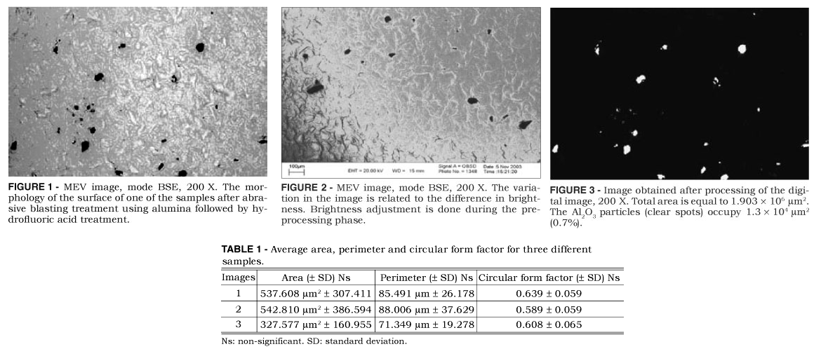
The surfaces treated with hydrofluoric acid-based chemicals showed morphological differences. Two cases of alumina-based blasted surfaces were studied: with and without posterior chemical attack using HF. After the chemical treatment, the surfaces showed a smoother, more homogeneous and more regular microtopography. Without the chemical attack, the surfaces showed irregularities, including peaks and depressions. Figure 1 shows the morphology of one of the samples after abrasive blasting treatment using alumina followed by hydrofluoric acid attack. The presence of small pits not larger than 10 micrometers in diameter may be observed throughout the surface of titanium. Although the acid treatment was effective in modifying the surface morphology and also in removing a large amount of aluminum from the blasting process, alumina residues were identified in the EDS analysis. These incrusted particles were also observed as darker regions in the MEV analysis (Figure 1: BSE mode – backscattered electrons).
The digital images of the surfaces were proc-essed using the KS 400 program (Carl Zeiss, Oberkochen, Germany) through a typical sequence of procedures related to analysis and digital proc-essing of images. The processing of images is based on mathematical operations capable of modifying the values of the image's pixels, with the objective of preparing them to be analyzed by the computer; in other words, identifying image attributes that can be related either to the manufacturing processes or to the material characteristics. The following procedures were followed in order to process and analyze the images: digital imaging (MEV); adjusting of the brightness and identification of the Al2O3 particles; after-processing (fulfilling of emptiness, corrections of the grain boundaries); definition of attributes that allowed measurement of the average area, perimeter, and circular form factor of the particles. Figure 2 shows a surface presenting variation of brightness in its diagonal axis, enhanced by a mask of colors. The variation in the image indicates a difference in the intensity of its pixels. Problems of brightness are corrected mathematically during an intermediate step in the image processing operations.
Form factors are used to identify specific forms in digital images. Normally, they are defined by a value that varies between 0 and 1, assuming a maximum value for objects with specific geometric forms, such as perfectly spherical forms. Specifically, circular form factors can be based on particle areas.
DISCUSSION
The role of surface contamination in implant failures is not yet well understood10. Since blasting processes are abrasive, they can not only modify the surface roughness13 but also remove contaminants resting on the metallic surface of the implant, making the surface more reactive16. It is possible that the use of abrasive particles whose chemical composition differs from the chemical composition of the implant may alter the biocompatibility of the implant's surface16. It has been suggested that the presence of inorganic contaminants could lead to lack of clinical success. Aluminum ions are suspected to impair bone formation by a possible competitive action with calcium1. Titanium implant casting associated with tissue breakdown has revealed embedded particles of alumina2.
Specific surface chemical treatments have been used for removing contaminants originating from either the implant's manufacturing proc-esses or the clinical surgical preparation. These treatments involve the use of solutions such as hydrofluoric acid (HF), sulfuric acid (H2SO4), nitric acid (HNO3), phosphoric acid (H3PO4), and organic acids, as well as methods utilizing anodic oxidation with electric current in acid environment and nitretation techniques5.
In the present research, digital image processing was used to quantify and create parameters related to the presence of residual Al2O3 on the surface of titanium blasted using aluminum oxide. The surfaces treated with hydrofluoric acid-based chemicals showed important morphological differences. After the chemical treatment, the surfaces showed a smoother, homogeneous and more regular microtopography, whereas without the chemical treatment the surfaces showed irregularities, including peaks and depressions. Although the acid treatment was effective in both modifying the surface morphology and in removing a large amount of aluminum from the blasting process4, alumina residues were identified in the EDS. These incrusted particles were also observed as darker regions in the MEV (BSE mode – backscattered electrons – Figures 1 and 2).
Figure 3 presents the image shown in Figure 1 after being processed and analyzed by KS 400 (Carl Zeiss, Oberkochen, Germany), which is a versatile image processing program designed to support demanding professional applications. The KS 400 program offers a wide range of different parameters for measurement and analysis. The area, perimeter and circular form of the Al2O3 particles were calculated digitally (Table 1). The results showed that the total contaminant particle area (depicted as clear regions in Figure 3) was of similar size for all samples. The area parameter of the individual particle showed higher dispersion of the distribution of values. For example, Al2O3 particles as large as 50 µm were mixed with very small ones, resulting in a high variation in the size of the particles (537.608 µm2 ± 307.411; 542.81 µm2 ± 386.594 and 327.577 µm2 ± 160.955, respectively, for the three images), and consequently in larger standard deviations. The mean area occupied by the aluminum oxide on the sample surfaces was 469.32 (± 284.98) µm2 (0.7% of the total area).
The perimeter parameter depends on both the morphology and the size of the particles of Al2O3. Consequently, results for the three processed images showed significant dispersion of measured values (85.491 µm ± 26.178; 88.006 µm ± 37.629 and 71.349 µm ± 19.278). The particle average perimeter was 81.61 µm.
The obtained values for the circular factor for the Al2O3 particles approximate a spherical form (0.639 ± 0.059; 0.589 ± 0.059 and 0.608 ± 0.065). The obtained circular factors are likely to be a common characteristic of all titanium surfaces blasted with Al2O3 particles, suggesting that the studied manufacturing process resulted in contaminated particles of regular and soft geometry, which tend to be spherical in form. The mean circular form factor was 0.60 (± 0.05).
Table 1 shows only the results related to the average values of area, perimeter and circular form factor related to the contaminant particles found in the three different samples. Differences in the measurements across distinct sample families were not calculated (analysis of variance). Although the results obtained in this study may not be extrapolated due to the small number of samples (or images) here utilized, we proposed a fast and accurate method for the analysis and processing of the digital images which may be employed in a considerable number of similar cases. The number of samples and images were reduced due to the high cost of the microscopy technology utilized in this study.
CONCLUSIONS
Using digital image processing, it was possible to accurately quantify and create parameters related to the presence of the Al2O3 phase, suggesting that this is an effective and appropriated process to characterize biomaterial-manufacturing processes. In spite of the difficulties related to the processing of digital images caused by the presence of "noises" as well as by low contrast level, the results obtained were significantly positive in characterizing the material.
Once a convenient sequence for the processing steps is defined, the sequence can be repeated for all images, creating a computational routine that makes for an easier processing of multiple images. After correctly performing the segmentation proc-ess, several attributes, such as the contaminant area, perimeter, principal dimensions, as well as form factor could be easily determined. Quantifying and characterizing the titanium surface is essential in both the evaluation of the material manufacturing process and its influence on the biological behavior of osteoblastic cell cultures.