Molecular Mechanism of Aluminum-Induced Oxidative Damage and Apoptosis in Rat Cardiomyocytes
Link: https://pubmed.ncbi.nlm.nih.gov/33634365/
LiuFang Zhou # 1, Mingjie He # 2, XiaoLan Li 3, Erbing Lin 4, YingChuan Wang 4, Hua Wei 4, Xi Wei 5
PMID: 33634365 DOI: 10.1007/s12011-021-02646-w
Abstract
Aluminum exposure can mediate either acute toxicity or chronic toxicity.
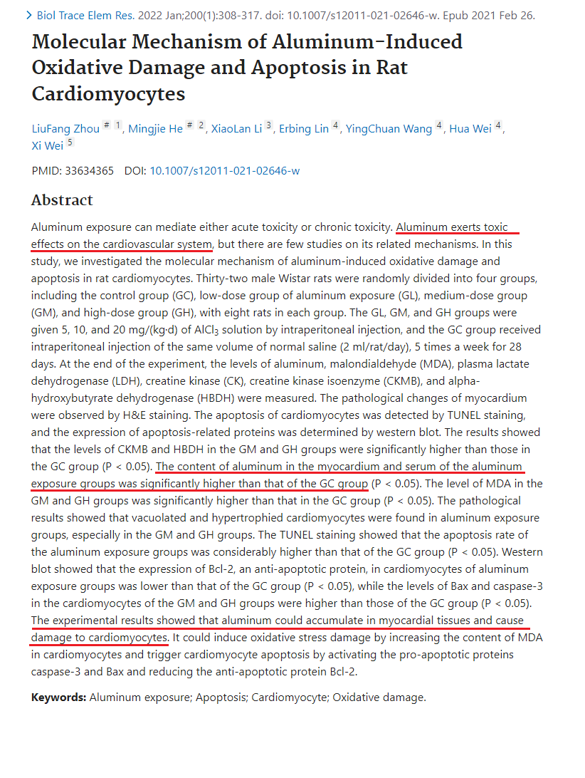
Aluminum exerts toxic effects on the cardiovascular system, but there are few studies on its related mechanisms.
In this study, we investigated the molecular mechanism of aluminum-induced oxidative damage and apoptosis in rat cardiomyocytes.
Thirty-two male Wistar rats were randomly divided into four groups, including the control group (GC), low-dose group of aluminum exposure (GL), medium-dose group (GM), and high-dose group (GH), with eight rats in each group. The GL, GM, and GH groups were given 5, 10, and 20 mg/(kg·d) of AlCl3 solution by intraperitoneal injection, and the GC group received intraperitoneal injection of the same volume of normal saline (2 ml/rat/day), 5 times a week for 28 days. At the end of the experiment, the levels of aluminum, malondialdehyde (MDA), plasma lactate dehydrogenase (LDH), creatine kinase (CK), creatine kinase isoenzyme (CKMB), and alpha-hydroxybutyrate dehydrogenase (HBDH) were measured. The pathological changes of myocardium were observed by H&E staining. The apoptosis of cardiomyocytes was detected by TUNEL staining, and the expression of apoptosis-related proteins was determined by western blot.
The results showed that the levels of CKMB and HBDH in the GM and GH groups were significantly higher than those in the GC group (P < 0.05). The content of aluminum in the myocardium and serum of the aluminum exposure groups was significantly higher than that of the GC group (P < 0.05). The level of MDA in the GM and GH groups was significantly higher than that in the GC group (P < 0.05). The pathological results showed that vacuolated and hypertrophied cardiomyocytes were found in aluminum exposure groups, especially in the GM and GH groups.
The TUNEL staining showed that the apoptosis rate of the aluminum exposure groups was considerably higher than that of the GC group (P < 0.05). Western blot showed that the expression of Bcl-2, an anti-apoptotic protein, in cardiomyocytes of aluminum exposure groups was lower than that of the GC group (P < 0.05), while the levels of Bax and caspase-3 in the cardiomyocytes of the GM and GH groups were higher than those of the GC group (P < 0.05).
The experimental results showed that aluminum could accumulate in myocardial tissues and cause damage to cardiomyocytes. It could induce oxidative stress damage by increasing the content of MDA in cardiomyocytes and trigger cardiomyocyte apoptosis by activating the pro-apoptotic proteins caspase-3 and Bax and reducing the anti-apoptotic protein Bcl-2.
Keywords: Aluminum exposure; Apoptosis; Cardiomyocyte; Oxidative damage.

See free reviews and follow our informative Blog to learn more: https://implante.institute/blog
See free analisys at: https://implante.institute/analises?perm_status=1
#implanteinstitute #implantcontamination #contamination #implantlost #titanium #zirconia #metal #aluminumfree #toxic #alumina #odontistry #materialscience #cellbiology #implantdentistry #dentist #implant #dental #dentalcare #plantcontentental #esthetic #cell
